Instituto de Investigação e Inovação em Saúde, Universidade do Porto
Rua Alfredo Allen, 208 4200-135 Porto, Portugal
Phone: +351 226 074 900
Email: info@i3s.up.pt
The NCN Lab combines experimental neurobiology, computational neuroscience and neuroengineering to understand how neural circuits process information and how their activity can be controlled. Our projects range from engineered neuronal circuits and brain-on-chip devices to adaptive neuromodulation strategies for neurological disease.
Neuronal population dynamics · engineered neuronal circuits · axonal electrophysiology · functional analysis · calcium imaging · microelectrode arrays · microfluidics · neural computation · microphysiological systems · human iPSC-derived neuronal cultures · closed-loop neuromodulation · neuromorphic computing · reinforcement learning · Parkinson’s disease · epilepsy · glioblastoma imaging.
We study how neuronal circuits generate, propagate and transform activity. By combining experimental recordings with computational analysis, we investigate how network architecture, cellular excitability and synaptic interactions shape information flow and population-level dynamics.
Microelectrode arrays are central to our work, enabling high-resolution recording and stimulation of neuronal populations. We also develop and validate neuro-electronic interfaces that support bidirectional communication with neural tissue and provide experimental platforms for closed-loop control.
We develop machine-learning approaches for analysing electrophysiological, imaging and clinical datasets. These tools support feature extraction, biomarker discovery, neural-state decoding, disease-state classification and the development of decision-support systems for neuromodulation.
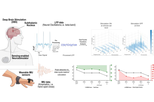
Through clinical collaborations, we analyse electrophysiological recordings from patients with neurological disorders. In Parkinson’s disease, we study sensing-enabled deep brain stimulation and biomarkers for adaptive DBS; in epilepsy, we investigate electrophysiological signatures that may support the identification and modulation of pathological neural activity.
We develop controlled microphysiological platforms that reproduce key aspects of neural organization in vitro. These systems integrate microfluidics, patterned neuronal cultures, imaging and electrophysiology to study circuit development, connectivity and perturbation responses under reproducible conditions.
We explore brain-inspired computational systems, including spiking neural networks and neuromorphic hardware, for real-time processing of neural signals. These approaches are particularly relevant for low-latency, energy-efficient neural interfaces and adaptive stimulation technologies.
We investigate how astrocytes respond to electrical stimulation and how neuron–astrocyte interactions shape network activity. By combining microelectrode arrays, functional imaging and engineered culture platforms, we aim to quantify astrocytic contributions to electrophysiological dynamics and neuromodulation.
We use primary, organotypic and human iPSC-derived neuronal cultures to monitor neural activity over time. These models allow us to investigate maturation, plasticity, disease-relevant phenotypes and the functional consequences of altered cellular composition or network structure.
We use computational models and simulations to interpret neural dynamics, test hypotheses and design stimulation strategies. Modelling allows us to connect mechanisms across scales, from cellular excitability and axonal propagation to network-level computation and control.
We design closed-loop strategies that use real-time neural recordings to guide stimulation. These approaches combine signal processing, control theory, reinforcement learning and neuro-electronic interfaces to modulate neuronal activity with precision and adaptivity.
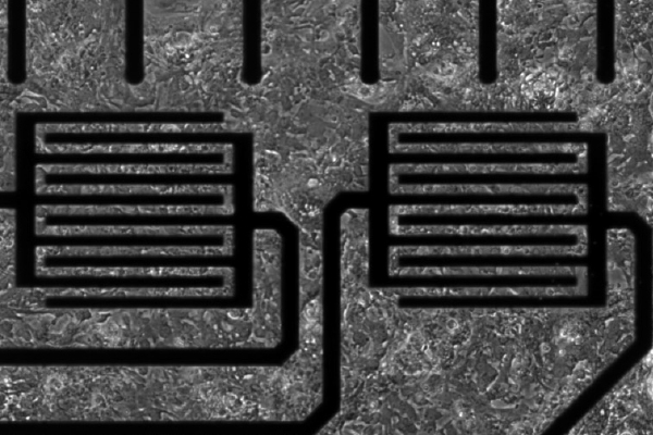
Neuron–astrocyte interactions are central to brain function, but their dynamics are difficult to quantify with conventional endpoint assays. We are developing microphysiological platforms that combine high-density microelectrode arrays, calcium imaging and microfluidics to monitor electrical and calcium signaling in neuron–astrocyte networks.
This project aims to generate standardized functional readouts of neuron–astrocyte communication at the network level. By validating the platform with known neuromodulators, we aim to create a robust tool for functional phenotyping and prediction of human-relevant neuroactive effects.
Suggested papers: astrocyte electrophysiology papers; microelectrode-array and microfluidic-platform papers; future neuron–astrocyte platform publication.
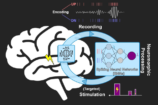
Closed-loop neuromodulation requires fast, efficient and adaptive computation. Neuromorphic computing offers a brain-inspired framework in which memory and processing are closely coupled, computation is event-based and parallel, and algorithms can be implemented with low latency.
In this project, we investigate spiking neural networks and neuromorphic hardware for real-time processing of neuronal recordings and targeted stimulation. The goal is to move toward adaptive neurotechnologies capable of detecting relevant electrophysiological states and modulating neural activity with high temporal precision.
Suggested papers: Dias et al., 2022, ACS Applied Electronic Materials; Castro et al., 2024, eLife; future NeuroSpark publications. The DBScope GitHub page also reflects the lab’s development of open computational tools for neural-device data analysis.
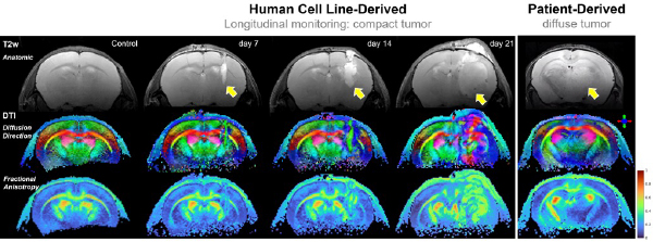
Glioblastoma (GBM) is the most aggressive primary brain tumor in adults and carries adismal prognosis. Therapeutic efficacy is limited by insufficient tools for individualized planning and response assessment, largely due to constraints of the current imaging standard, magnetic resonance imaging (MRI). Conventional MRI cannot reliably delineate the infiltrative tumor rim and lacks specificity to distinguish true- versus pseudo-progression. Advanced diffusion MRI enables in vivo quantitative characterization of tissue microstructure, with applications in the healthy brain, stroke, and neurodegeneration. By integrating mouse models of human GBM, advanced diffusion encoding and compartment modeling, translational 3 Tesla MRI, and histopathological validation, we aim to develop animaging approach for in vivo microstructural characterization of GBM.
Suggested papers: Simões et al., 2025, eLife; diffusion MRI and tumor-imaging publications from Rui Simões’ work.
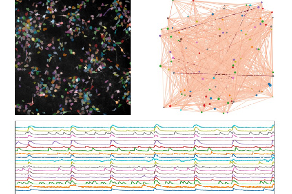
We are establishing in vitro models of neurodevelopment using human induced pluripotent stem cell (iPSC)-derived neuronal networks integrated with microelectrode arrays, calcium imaging, and microfluidic platforms. Through this combination, we capture the emergence of activity patterns and functional connectivity across development.
We engineer controlled microenvironments to dissect how cellular composition, network architecture, and activity-dependent mechanisms shape circuit maturation. These approaches enable longitudinal, high-resolution measurements of human neural dynamics, addressing intrinsic maturation timelines of human neurons.
By linking multiscale functional phenotypes to developmental trajectories and disease-relevant perturbations, we aim to uncover mechanisms underlying neurodevelopmental disorders and provide robust platforms for human-relevant functional screening.
Glioblastoma (GBM) is the most aggressive primary brain tumor in adults and carries adismal prognosis. Therapeutic efficacy is limited by insufficient tools for individualized planning and response assessment, largely due to constraints of the current imaging standard, magnetic resonance imaging (MRI). Conventional MRI cannot reliably delineate the infiltrative tumor rim and lacks specificity to distinguish true- versus pseudo-progression. Advanced diffusion MRI enables in vivo quantitative characterization of tissue microstructure, with applications in the healthy brain, stroke, and neurodegeneration. By integrating mouse models of human GBM, advanced diffusion encoding and compartment modeling, translational 3 Tesla MRI, and histopathological validation, we aim to develop an imaging approach for in vivo microstructural characterization of GBM.
Suggested papers: Simões et al., 2025, eLife; diffusion MRI and tumor-imaging publications from Rui Simões’ work.